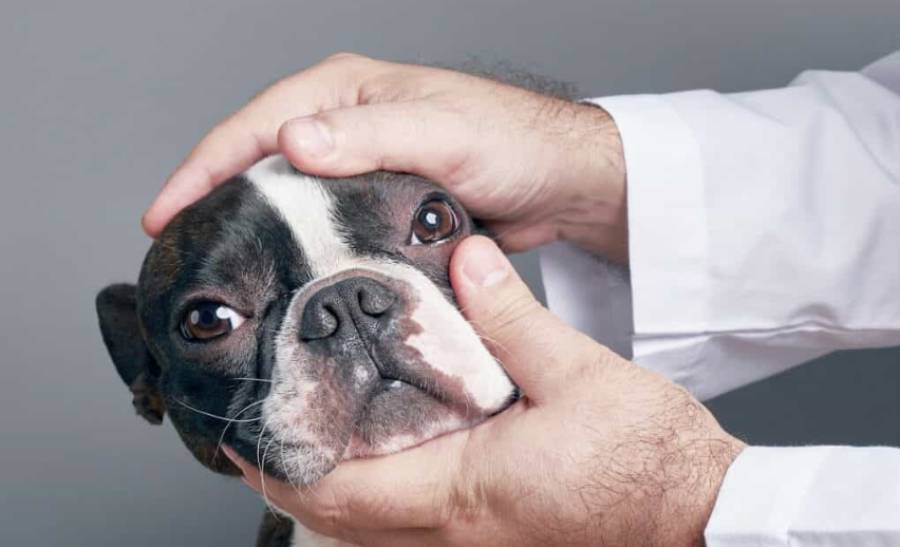
Boston Terrier corneal ulcers are painful injuries to the clear outer surface of the eye that require prompt veterinary attention. Boston Terriers can be particularly vulnerable because their prominent eyes are more exposed to scratches, foreign material, and other forms of trauma than eyes positioned deeper within the skull. Problems affecting the tear film, eyelids, or eyelashes can further increase the risk of corneal damage.
A Boston Terrier with a corneal ulcer may squint, keep one eye closed, produce excessive tears, develop redness or discharge, or repeatedly paw at the affected eye. The cornea may also appear cloudy or visibly abnormal in more noticeable cases. However, an ulcer is not always easy to see without an eye examination, so the absence of an obvious wound does not rule out corneal injury.
Corneal ulcers vary in depth and severity. A superficial ulcer may heal relatively quickly with appropriate veterinary treatment, while a deeper, infected, or complicated ulcer can damage more of the cornea and threaten vision. A veterinarian can examine the eye and use tests such as fluorescein staining to identify corneal damage. Treatment then depends on the ulcer’s depth, underlying cause, infection status, and healing response. This guide explains the causes, symptoms, diagnosis, treatment, recovery, complications, recurrence, and prevention of corneal ulcers in Boston Terriers.
What Is a Corneal Ulcer in a Boston Terrier?
A corneal ulcer in a Boston Terrier is an open sore or defect in the cornea, the transparent tissue covering the front of the eye. The cornea protects the internal structures of the eye while allowing light to enter. When part of its outer protective layer is damaged, the underlying corneal tissue becomes exposed, producing an ulcer that can cause significant pain and inflammation.
Corneal ulcers differ in depth. A superficial ulcer affects the outer layers of the cornea, while a deeper ulcer extends farther into the corneal tissue. This distinction matters because deeper damage carries a greater risk of complications, including infection, scarring, corneal perforation, and impaired vision. An ulcer can also become more complicated when an underlying eye problem prevents normal healing.
Corneal damage is particularly painful because the cornea is highly sensitive. A Boston Terrier with an ulcer may therefore react strongly even when the affected area appears small. Squinting, excessive tearing, keeping the affected eye closed, and rubbing at the face are common responses to this discomfort. The size or visibility of an eye injury should not be used by an owner to determine its severity because veterinary examination is needed to establish the location and depth of corneal damage.
Why Are Boston Terriers Prone to Corneal Ulcers?
Boston Terriers are prone to corneal ulcers partly because their brachycephalic head shape leaves their prominent eyes relatively exposed to injury and irritation. Unlike an eye positioned deeper within the socket, a prominent eye has greater exposure to the surrounding environment. This increases opportunities for the cornea to contact objects that can scratch or irritate its surface.
Everyday activities can create opportunities for corneal trauma. A Boston Terrier can injure the cornea by running through vegetation, contacting a branch or other object, playing roughly with another animal, or scratching an irritated eye with a paw. Small foreign materials that reach the ocular surface can also produce irritation and subsequent corneal injury. The breed’s exposed eyes make monitoring these seemingly minor incidents particularly important.
Protection of the cornea also depends on more than eye position. A healthy tear film, normal eyelids, and correctly positioned eyelashes help maintain and protect the ocular surface. Tears lubricate the cornea and help prevent excessive dryness, while normal blinking distributes the tear film across its surface. Problems that interfere with these protective mechanisms can leave the cornea more susceptible to injury or interfere with healing after an ulcer develops.
Eyelid and eyelash abnormalities can create another source of repeated irritation when they cause hair or tissue to contact the corneal surface. Similarly, inadequate tear production can reduce lubrication and protection. These factors do not mean every Boston Terrier will develop a corneal ulcer, but they help explain why eye discomfort in this breed deserves attention rather than being treated as a minor cosmetic problem.
What Causes Corneal Ulcers in Boston Terriers?
Corneal ulcers in Boston Terriers can develop when trauma, foreign material, inadequate lubrication, eyelid or eyelash abnormalities, or another ocular problem damages the corneal surface. These causes differ in origin, but each can disrupt the protective outer layer of the cornea and expose the tissue underneath.
Physical trauma is a direct cause of corneal injury. A Boston Terrier can scratch its cornea while moving through bushes or other vegetation, playing with another animal, or contacting furniture and other objects. The dog’s own paw can also damage the cornea when eye irritation causes repeated scratching or rubbing. Because Boston Terriers have prominent eyes, their corneas have greater exposure to these environmental hazards.
Foreign material can damage the cornea through direct contact or continued irritation. Small particles, plant material, hair, and similar debris can become trapped against the ocular surface. The material may scratch the cornea directly or trigger rubbing that causes additional trauma. Persistent squinting, tearing, or pawing after outdoor activity therefore warrants attention even when the owner cannot see anything inside the eye.
Inadequate lubrication can make the corneal surface more vulnerable. The tear film normally lubricates the eye and supports a healthy ocular surface. When tear production or tear-film function is impaired, the cornea loses part of this protection. A poorly lubricated surface is more susceptible to irritation and injury and may also have difficulty healing normally after an ulcer develops.
Eyelid and eyelash abnormalities can cause repeated mechanical irritation. Hair or abnormal tissue that repeatedly contacts the cornea can damage its surface over time. In these cases, treating the ulcer alone may not address the entire problem because the source of repeated corneal contact can remain. A veterinarian may therefore investigate an underlying ocular abnormality when an ulcer recurs or does not heal as expected.
What Are the Symptoms of a Corneal Ulcer in a Boston Terrier?
The main signs of a corneal ulcer in a Boston Terrier include squinting, keeping the affected eye closed, excessive tearing, redness, discharge, pawing at the eye, and changes in the appearance of the cornea. These signs reflect pain, inflammation, or irritation of the ocular surface, but they cannot confirm a corneal ulcer without a veterinary eye examination.
Squinting is an important sign because corneal ulcers can be painful. A Boston Terrier may partially close the affected eye or refuse to open it normally. Some dogs become more sensitive to light or turn their face away when the eye is examined. A sudden change from normally open eyes to persistent squinting or closure of one eye should not be dismissed simply because no wound is visible.
Excessive tearing can occur as the eye responds to corneal irritation. Owners may notice moisture around the eye or tears running down the face. Redness can develop at the same time because ocular irritation triggers an inflammatory response. Eye discharge may also appear, although its presence and appearance alone do not establish the cause of the problem.
Pawing and rubbing are behavioral signs of discomfort. A Boston Terrier may repeatedly touch the affected eye with a paw or rub the side of its face against furniture, carpet, or bedding. This behavior matters for two reasons: it signals that the eye is uncomfortable, and repeated rubbing can produce additional trauma to an already damaged cornea.
Visible corneal changes can occur in more noticeable or advanced cases. The normally transparent cornea may appear cloudy, hazy, discolored, or otherwise abnormal. However, a corneal ulcer does not have to be visible to the owner. Small or superficial defects can require fluorescein staining and a veterinary eye examination to identify. Persistent squinting, tearing, redness, or eye pain therefore warrants veterinary assessment even when the cornea appears normal at home.
Is a Corneal Ulcer in a Boston Terrier an Emergency?
A suspected corneal ulcer in a Boston Terrier requires prompt veterinary attention because corneal damage can worsen and potentially threaten the eye if treatment is delayed. The urgency increases when the dog has severe pain, keeps the eye closed, develops pronounced cloudiness or discharge, has an obvious eye injury, or shows a rapid change in the appearance of the eye.
Corneal ulcers are not all equally severe. A superficial injury involves the outer portion of the cornea, while deeper damage extends farther into the corneal tissue. Owners cannot reliably determine this depth by looking at the eye at home. A small visible defect can still be painful, and some clinically important corneal injuries may not be clearly visible without diagnostic testing.
Persistent squinting or keeping one eye closed is a particularly important reason to seek veterinary care. These behaviors indicate ocular discomfort and can occur when the sensitive corneal surface is injured. Pronounced cloudiness, worsening redness, abnormal discharge, visible corneal changes, or an obvious traumatic event add further concern. Changes suggesting impaired vision or rapidly worsening eye pain also require urgent assessment.
Delayed treatment can allow an existing ulcer to become more complicated. Continued rubbing may produce additional trauma, while infection or an unresolved underlying eye problem can interfere with normal healing. Deeper corneal damage carries more serious consequences, including scarring, perforation, and loss of vision in severe cases. Prompt examination allows the veterinarian to determine whether an ulcer is present and select treatment according to its severity and underlying cause.
How Does a Veterinarian Diagnose a Corneal Ulcer in a Boston Terrier?
A veterinarian diagnoses a corneal ulcer by examining the Boston Terrier’s eye and using diagnostic tests, particularly fluorescein staining, to identify damage to the corneal surface. The examination also helps determine the severity of the injury and whether another ocular problem contributed to the ulcer.
The veterinarian first evaluates the eye and surrounding structures for abnormalities. This assessment can include the cornea, eyelids, eyelashes, ocular surface, and visible signs of inflammation or trauma. Examining these structures matters because an ulcer may result from more than a single accidental scratch. Persistent eyelid, eyelash, tear-film, or ocular surface problems can continue irritating the cornea after the initial injury.
Fluorescein staining is commonly used to identify corneal surface defects. A fluorescein dye is applied to the eye and adheres to areas where the normal corneal surface has been disrupted. The resulting staining pattern helps the veterinarian locate the defect and evaluate characteristics of the damaged area that cannot be assessed reliably by an owner looking at the eye at home.
Diagnosis does not end with confirming that an ulcer exists. The veterinarian also evaluates its depth, appearance, progression, and possible underlying cause because these factors influence treatment and prognosis. Additional testing may be appropriate when tear production, infection, recurrent injury, or another ocular disorder is suspected.
Follow-up examinations can also form an important part of the diagnostic process. A Boston Terrier may appear more comfortable after treatment begins even though the cornea has not completely healed. Re-examining the eye and repeating diagnostic testing when appropriate allows the veterinarian to confirm healing rather than relying solely on improvement in visible symptoms.
How Are Corneal Ulcers in Boston Terriers Treated?
Corneal ulcers in Boston Terriers are treated according to the depth of the ulcer, its underlying cause, the presence of infection, and how well the cornea is healing. Treatment can include veterinary ophthalmic medications, pain management, protection from further eye trauma, correction of an underlying problem, and surgical treatment for deeper or complicated ulcers.
A superficial, uncomplicated ulcer generally requires a different treatment approach from a deep or non-healing ulcer. The veterinarian therefore determines the condition of the cornea before selecting treatment. An ulcer caused by a one-time scratch may require management of the corneal injury itself, while recurrent damage associated with an eyelid, eyelash, tear-film, or other ocular problem may require treatment of both the ulcer and its underlying cause.
Preventing additional trauma is also an important part of treatment. Eye pain and irritation can cause a Boston Terrier to paw at its face or rub the affected eye against furniture, bedding, or carpet. This behavior can further damage the cornea and interfere with healing. An Elizabethan collar may therefore be used to prevent the dog from reaching the affected eye while the cornea recovers.
Owners should use ophthalmic medications only as directed by a veterinarian. Eye problems that appear similar can have different causes, and a product that is appropriate for one ocular condition is not automatically appropriate for a corneal ulcer. Veterinary examination is particularly important before using previously prescribed or leftover eye medication.
What Medications Are Used for Boston Terrier Corneal Ulcers?
Veterinarians select medications for Boston Terrier corneal ulcers based on the ulcer’s characteristics and the problems that need to be controlled during healing. Depending on the individual case, treatment may include ophthalmic medication intended to manage infection risk or infection, medications for pain, and additional therapy directed at an underlying ocular condition.
Antibiotic eye drops or ointments may be prescribed when the veterinarian determines they are appropriate. Damage to the normal corneal surface compromises part of the eye’s protective barrier, so preventing or controlling bacterial complications can be an important treatment objective. The specific medication and frequency of administration depend on the clinical findings rather than on the dog’s breed alone.
Pain management is another component of treatment because corneal injuries can cause substantial discomfort. Reducing pain improves the dog’s comfort and can help reduce behaviors associated with ocular irritation. Pain relief does not confirm that the ulcer has healed, however. A Boston Terrier that begins opening its eye normally may still require the complete prescribed treatment and a veterinary recheck.
Treatment may also address conditions that contributed to the ulcer. For example, a problem affecting lubrication or repeated physical irritation of the cornea can continue interfering with recovery unless it is identified and managed. This is why treatment of a recurrent or poorly healing ulcer can involve more than applying medication to the damaged corneal surface.
When Does a Boston Terrier Corneal Ulcer Need Surgery?
A Boston Terrier may need a surgical or ophthalmic procedure when a corneal ulcer is deep, complicated, at risk of threatening the integrity of the eye, or fails to heal normally with appropriate medical management. The decision depends on examination findings and should be made by a veterinarian or veterinary ophthalmologist.
Deep ulcers require particular attention because progressive loss of corneal tissue can weaken the structure of the eye. Severe damage can eventually lead to corneal perforation, which places vision and the eye itself at risk. In these situations, treatment may need to do more than control discomfort or infection; the damaged cornea may require additional support or specialized intervention.
A procedure may also be considered when an ulcer repeatedly fails to heal as expected. In these cases, the veterinarian investigates why normal corneal repair is not occurring and selects an intervention based on the type of lesion and underlying problem. Referral to a veterinary ophthalmologist may be appropriate for complex cases requiring specialized evaluation or treatment.
Medical or surgical treatment should be followed by appropriate rechecks. Improved redness, reduced tearing, or less squinting does not by itself prove that the corneal defect has completely healed. Veterinary reassessment allows healing to be confirmed and treatment to be adjusted or discontinued at the appropriate stage.
How Long Does a Boston Terrier Corneal Ulcer Take to Heal?
The healing time for a Boston Terrier corneal ulcer depends on its depth, underlying cause, infection status, continued irritation, and response to treatment. A superficial uncomplicated ulcer can heal faster than a deep, infected, recurrent, or otherwise complicated ulcer. For this reason, the appearance of the eye alone cannot provide a reliable healing timeline.
Corneal healing requires the damaged surface to repair while the eye is protected from further injury. Continued rubbing, repeated contact from an eyelid or eyelash abnormality, inadequate ocular surface protection, or another unresolved problem can interfere with this process. Treatment adherence also matters because prescribed eye medications and protective measures are intended to create conditions that allow the cornea to recover without additional damage.
Improvement in symptoms does not necessarily mean the ulcer has completely healed. A Boston Terrier may squint less, produce fewer tears, or appear more comfortable before the corneal defect has fully resolved. Stopping treatment solely because the eye looks better can therefore be inappropriate. Owners should follow the treatment schedule and recheck instructions provided by their veterinarian.
Veterinary follow-up provides a more reliable way to assess recovery. The veterinarian can re-examine the cornea and repeat fluorescein staining when appropriate to determine whether the surface defect remains. An ulcer that is not healing as expected may require further investigation for infection, repeated trauma, an ocular surface disorder, or another underlying factor that is delaying normal corneal repair.
What Complications Can Corneal Ulcers Cause in Boston Terriers?
Corneal ulcers in Boston Terriers can lead to persistent corneal damage, infection, scarring, deeper tissue loss, corneal perforation, and vision impairment when the injury becomes complicated or progresses. The risk depends on the depth and cause of the ulcer, whether infection is present, and how quickly effective treatment begins.
A non-healing ulcer is one important complication because persistent damage leaves the corneal surface vulnerable for longer. An ulcer may fail to heal normally when the original cause remains present or another ocular condition interferes with repair. Repeated rubbing or continued mechanical irritation can also damage newly healing tissue and prolong the problem.
Infection can make corneal damage more serious. The intact corneal surface normally provides a protective barrier, but an ulcer disrupts that barrier. Bacterial involvement can contribute to inflammation and tissue damage, making veterinary monitoring important when the appearance or symptoms of an ulcer worsen rather than improve.
Corneal scarring can occur after tissue damage and healing. Because the cornea needs to remain transparent for light to pass efficiently into the eye, substantial scarring can affect vision depending on its location and severity. The clinical significance of a scar therefore depends on more than whether visible discoloration remains after the ulcer heals.
Deep progression creates the greatest structural concern. As an ulcer extends farther through the cornea, the remaining tissue becomes thinner and weaker. Severe tissue loss can ultimately result in corneal perforation, compromising the integrity of the eye and potentially threatening vision. This is one reason a painful or visibly abnormal eye should be examined promptly rather than monitored at home to see whether it improves.
Vision impairment is a potential consequence of severe corneal disease rather than an inevitable result of every ulcer. Prompt diagnosis, protection of the injured eye, appropriate treatment, and veterinary rechecks reduce the opportunity for a manageable corneal injury to progress unnoticed. Owners should therefore treat worsening pain, increasing cloudiness, marked discharge, or significant changes in the appearance of the eye as reasons for veterinary reassessment.
Can Corneal Ulcers Come Back in Boston Terriers?
Corneal ulcers can recur in Boston Terriers when the factor that damaged the cornea remains present or the eye experiences another injury. Recurrent ulcers are particularly important to investigate because healing the current corneal defect does not necessarily eliminate the condition that allowed it to develop.
Repeated physical trauma is one possible cause of recurrence. Boston Terriers have prominent eyes that remain relatively exposed during normal activities, so contact with vegetation, rough play, foreign material, or scratching can produce another corneal injury after a previous ulcer has healed. Repeated pawing or rubbing caused by persistent eye irritation can create the same problem.
An eyelid or eyelash abnormality can also expose the cornea to repeated mechanical irritation. When hair or another structure repeatedly contacts the ocular surface, treating the resulting ulcer addresses the damaged tissue but not necessarily the source of the contact. Identifying and managing the underlying abnormality can therefore be an important part of preventing another episode.
Problems affecting the tear film or ocular surface can contribute to recurrence as well. Adequate lubrication helps protect the cornea from friction and environmental irritation. When this protective function is impaired, the ocular surface can remain vulnerable even after an existing ulcer closes.
A Boston Terrier that develops repeated ulcers should therefore receive veterinary evaluation rather than having each episode treated as an unrelated injury. The clinical objective is to heal the current ulcer and identify persistent factors that could cause another one. A veterinarian may evaluate the eyelids, eyelashes, tear production, ocular surface, and other structures based on the dog’s examination findings and history.
How Can You Reduce the Risk of Corneal Ulcers in a Boston Terrier?
You can reduce a Boston Terrier’s risk of corneal ulcers by limiting avoidable eye trauma, monitoring the eyes for early signs of irritation, preventing persistent rubbing, and managing underlying ocular problems with veterinary care. Prevention cannot eliminate every corneal injury, particularly in a breed with prominent eyes, but it can reduce preventable exposure and help problems receive attention before additional damage occurs.
Monitor both eyes regularly for changes in appearance and behavior. Squinting, excessive tearing, redness, discharge, repeated blinking, or pawing at an eye can indicate ocular discomfort even when no injury is visible. Recognizing these changes early is important because a small corneal defect may not be apparent without veterinary examination.
Reduce exposure to obvious sources of eye trauma when practical. Dense vegetation, thorny plants, sharp branches, and situations involving uncontrolled rough contact can place prominent eyes at greater risk. This does not mean a Boston Terrier needs to avoid normal exercise or outdoor activity. Instead, owners can pay particular attention to environments where objects are likely to contact the dog’s face and eyes.
Prevent persistent rubbing when an eye is already irritated. Pawing at the face or rubbing the eye against furniture and flooring can turn irritation into additional corneal trauma or worsen an existing injury. A veterinarian may recommend an Elizabethan collar when physical protection is needed during treatment or recovery.
Manage diagnosed eye conditions according to veterinary instructions. Problems involving tear production, eyelids, eyelashes, or other parts of the ocular surface can increase corneal exposure or irritation when they remain unresolved. Addressing these conditions targets a risk factor rather than repeatedly treating the resulting corneal damage.
Regular observation is particularly valuable for Boston Terriers because their eye conformation makes visible changes relatively easy for owners to notice. The practical goal is not to prevent every possible corneal ulcer but to minimize avoidable trauma and respond promptly when signs of eye pain or irritation appear.
Read more: Boston Terrier Cherry Eye: Symptoms, Causes & Treatment
What Other Eye Problems Can Look Like a Corneal Ulcer in a Boston Terrier?
Several eye problems can produce signs similar to a corneal ulcer in a Boston Terrier, including dry eye, conjunctival inflammation, foreign material in the eye, and other painful corneal or ocular conditions. Squinting, redness, tearing, discharge, and rubbing indicate that an eye is irritated or painful, but these signs alone do not identify the underlying condition.
Dry eye can produce ocular irritation when the tear film does not adequately protect and lubricate the surface of the eye. Affected dogs may develop redness, discomfort, discharge, or changes to the corneal surface. Dry eye can also matter beyond symptom overlap because inadequate ocular surface protection can contribute to corneal damage.
Conjunctival inflammation can also cause redness, tearing, discharge, and irritation. These visible signs can resemble those associated with a corneal injury, particularly when an owner is examining the eye without veterinary equipment. The presence of redness or discharge therefore does not establish whether the primary problem involves the conjunctiva, cornea, or another ocular structure.
Foreign material can produce another similar presentation. Plant fragments, hair, debris, or other material contacting the eye can cause sudden squinting, tearing, and pawing. Foreign material may also damage the corneal surface itself, meaning a foreign body and a corneal ulcer can occur together rather than being mutually exclusive conditions.
Other painful ocular disorders can produce similar behavioral signs because dogs have a limited number of ways to express eye discomfort. A Boston Terrier may close the eye, blink repeatedly, avoid bright light, or rub its face regardless of the exact structure causing the pain. A veterinary eye examination is therefore needed to distinguish a corneal ulcer from other conditions that produce similar symptoms.
What Should You Do If You Think Your Boston Terrier Has a Corneal Ulcer?
Contact a veterinarian promptly if your Boston Terrier develops persistent squinting, keeps an eye closed, has excessive tearing or discharge, repeatedly rubs the eye, or develops visible corneal changes. Owners cannot reliably determine the depth or severity of a corneal injury at home, and some ulcers are difficult to see without diagnostic staining.
Prevent additional rubbing while arranging veterinary care. Pawing at the eye or rubbing the face against furniture, carpet, or bedding can cause further corneal trauma. Physical protection, including an Elizabethan collar when appropriate and available, can help prevent self-inflicted damage while the dog is being evaluated or during treatment.
Do not use leftover or previously prescribed ophthalmic medication unless a veterinarian specifically instructs you to use it for the current problem. Similar-looking eye conditions can require different treatment approaches. Confirming the diagnosis before treatment also allows the veterinarian to assess the ulcer’s depth, investigate contributing factors, and determine whether infection or another ocular problem is present.
Follow the complete treatment and recheck plan after diagnosis. Improvement in squinting, tearing, redness, or discomfort can occur before the corneal surface has fully healed. Veterinary reassessment, including repeat fluorescein staining when indicated, provides a more reliable way to confirm that the defect has resolved.
The practical response follows a clear sequence: recognize signs of eye pain, prevent additional trauma, obtain veterinary diagnosis, follow prescribed treatment, and complete recommended rechecks. This approach addresses both the immediate corneal injury and any underlying problem that could delay healing or contribute to another ulcer.
2 thoughts on “Boston Terrier Corneal Ulcers: Symptoms & Treatment”