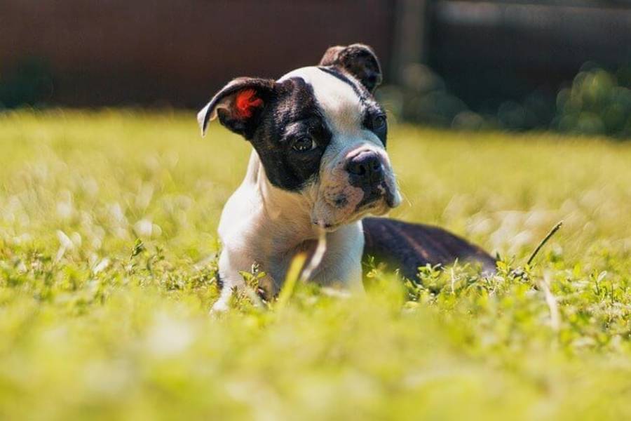
Boston Terrier cherry eye is a prolapse of the gland associated with the third eyelid, which usually appears as a pink or red rounded mass at the inner corner of the eye. The condition develops when the tissue that normally holds the gland in position no longer provides sufficient support, allowing the gland to protrude and become visible. Because this gland contributes to the tear film that protects the surface of the eye, cherry eye is more than a cosmetic change.
Boston Terriers can develop cherry eye in one eye or both eyes, and owners may also notice redness, swelling, excessive tearing, discharge, squinting, or rubbing at the affected eye. A visible red mass near the inner corner is a characteristic sign, but similar-looking eye problems can occur. A veterinary examination is therefore important for confirming that the protruding tissue is the third eyelid gland and assessing the rest of the eye.
Treatment depends on the condition of the gland and the severity of the prolapse. Some cases receive supportive medical treatment, while persistent cherry eye may require surgery to reposition and preserve the gland. This guide explains the symptoms and causes of Boston Terrier cherry eye, its potential complications, treatment options, surgery, recovery, recurrence, and prevention.
What Is Cherry Eye in a Boston Terrier?
Cherry eye in a Boston Terrier is the visible prolapse of the gland associated with the third eyelid. Instead of remaining in its normal position near the inner corner of the eye, the gland moves outward and becomes visible as a pink or red mass. The medical problem involves the gland and the tissues that normally hold it in place, rather than the eyeball itself.
Dogs have a third eyelid, also called the nictitating membrane, in addition to the upper and lower eyelids. This structure sits at the inner corner of the eye and helps protect the ocular surface. Associated with the third eyelid is a gland that contributes to the tear film covering and lubricating the eye. When the supporting tissue fails to keep this gland in its normal position, the gland can protrude above the edge of the third eyelid.
The exposed gland produces the appearance responsible for the name “cherry eye.” The protruding tissue is usually rounded, pink, or red and can resemble a small cherry at the inner corner of the eye. Exposure can also cause the gland to become swollen and irritated, making it appear larger or redder than it did when the prolapse first developed.
The gland’s role in tear production is one reason cherry eye should not be treated only as a cosmetic problem. Maintaining a stable tear film helps lubricate and protect the cornea and other tissues on the surface of the eye. Treatment therefore focuses not only on improving the appearance of the eye but also on maintaining the function of the affected gland whenever possible.
What Does Cherry Eye Look Like in a Boston Terrier?
Cherry eye usually looks like a rounded pink or red lump protruding from the inner corner of a Boston Terrier’s eye. The mass can be relatively small or become more prominent as the exposed gland becomes swollen and inflamed. Its position near the nose-facing corner of the eye is one of the most recognizable characteristics of the condition.
A Boston Terrier with cherry eye may have additional signs around the affected eye. These can include redness, swelling, increased tearing, eye discharge, squinting, and repeated rubbing or pawing at the face. These signs do not occur with the same severity in every dog because the degree of irritation depends on the condition of the exposed gland and surrounding ocular tissues.
Cherry eye can affect one eye or both eyes. A Boston Terrier may initially develop a visible prolapse in only one eye, so a normal-looking second eye does not rule out cherry eye in the affected eye. Owners should monitor both eyes for changes rather than assuming that the condition must appear bilaterally.
A red or swollen structure in the inner corner of the eye should not automatically be diagnosed as cherry eye at home. Eye inflammation, conjunctival problems, trauma, and other ocular disorders can also cause redness, swelling, discharge, or discomfort. The location and characteristic appearance of a prolapsed third eyelid gland provide useful clues, but a veterinarian can examine the eye and determine which structure is actually affected.
Early veterinary assessment is particularly important when the Boston Terrier is squinting, repeatedly rubbing the eye, producing abnormal discharge, or showing a noticeable change in the surface of the eye. These signs indicate that the problem involves more than appearance and that the eye needs to be evaluated for irritation or another concurrent condition.
Why Do Boston Terriers Get Cherry Eye?
Boston Terriers develop cherry eye when the tissue that normally holds the third eyelid gland in position fails to provide enough support. The weakened attachment allows the gland to move out of its normal location and protrude above the third eyelid. Once exposed, the gland can become swollen and inflamed, producing the characteristic red or pink mass at the inner corner of the eye.
The underlying mechanism can be described as a sequence: supporting tissue becomes insufficient, the third eyelid gland shifts from its normal position, the gland protrudes, and the exposed tissue becomes increasingly visible. This explains why cherry eye is fundamentally a structural problem rather than simply eye redness or temporary inflammation. Inflammation can make an existing prolapse more noticeable, but reducing redness alone does not necessarily correct the displaced gland.
Breed predisposition also contributes to cherry eye risk. Boston Terriers are among the dog breeds in which owners and veterinarians may encounter prolapse of the third eyelid gland. A predisposition means the anatomical support surrounding the gland is more likely to allow prolapse; it does not mean every Boston Terrier will develop the condition. Two dogs of the same breed can therefore have different outcomes even when they receive similar routine eye care.
Cherry eye is also commonly associated with younger dogs, making a newly developed mass at the inner corner of a young Boston Terrier’s eye particularly important to examine. However, age alone cannot confirm the condition. The appearance and location of the tissue, accompanying eye changes, and veterinary examination are more useful for identifying a prolapsed third eyelid gland.
Owners generally cannot prevent the underlying structural weakness through routine cleaning, diet, or other basic care. This distinction matters because cherry eye should not automatically be interpreted as evidence of poor care. The relevant problem is the gland’s loss of normal structural support, so treatment decisions focus on the affected gland and surrounding tissues rather than attempting to correct an assumed hygiene problem.
Is Cherry Eye Painful for a Boston Terrier?
Cherry eye is not necessarily severely painful by itself, but an exposed and inflamed third eyelid gland can cause significant irritation and discomfort in a Boston Terrier. The degree of discomfort varies according to the amount of swelling, inflammation, dryness, and irritation affecting the exposed tissue and surrounding eye.
A prolapsed gland is no longer protected in its normal anatomical position. Exposure can irritate its surface and surrounding tissues, leading to redness and swelling. As irritation increases, the Boston Terrier may squint, blink more frequently, rub its face against objects, or use a paw to scratch around the affected eye. Increased tearing or discharge may occur at the same time.
Behavior provides useful information about discomfort. A Boston Terrier that repeatedly paws at the eye, keeps the eyelids partly closed, resists having the face touched, or suddenly becomes more protective of the affected side should receive veterinary attention. Persistent rubbing is particularly important because mechanical contact can further irritate an already inflamed eye.
Visible discomfort should also not automatically be attributed to cherry eye alone. Pronounced squinting or apparent eye pain can accompany other ocular problems, including conditions affecting the cornea. A veterinarian can examine the protruding gland and the rest of the eye to determine whether cherry eye is the only problem present.
Owners should therefore evaluate cherry eye based on both the visible mass and the Boston Terrier’s behavior. A relatively calm dog with a small prolapse still needs appropriate assessment, while obvious pain, persistent squinting, repeated rubbing, or substantial discharge increases the importance of prompt veterinary evaluation.
Is Cherry Eye Dangerous for a Boston Terrier?
Cherry eye is usually not a life-threatening condition, but leaving a prolapsed third eyelid gland untreated can increase the risk of persistent irritation, inflammation, and problems with the tear film. The main concern is preserving the health and function of the affected gland rather than the appearance of the red mass alone.
The third eyelid gland contributes to the tears that keep the ocular surface lubricated. When the gland remains prolapsed, its normal position and function are disrupted, while the exposed tissue is more vulnerable to irritation. Persistent exposure can cause the gland to become increasingly swollen and inflamed, making the prolapse more noticeable and potentially more uncomfortable for the Boston Terrier.
An untreated prolapse can also create longer-term concerns about tear production and the condition of the ocular surface. Tears provide lubrication and help maintain a healthy environment over the cornea and conjunctiva. For this reason, preserving a functional third eyelid gland is an important treatment objective when a veterinarian manages cherry eye.
The urgency of veterinary attention also depends on the signs accompanying the prolapse. A Boston Terrier should receive prompt veterinary evaluation when the eye becomes markedly red or swollen, develops abnormal discharge, remains partly closed, or causes repeated pawing and rubbing. A cloudy or otherwise abnormal-looking corneal surface also warrants veterinary attention because another eye disorder may be present alongside the prolapsed gland.
Waiting for a persistent cherry eye to disappear without assessment is not a reliable treatment strategy. Even when the protruding tissue temporarily becomes less visible, the structural weakness that allowed the gland to move may remain. Veterinary assessment helps determine the condition of the gland, identify additional eye problems, and establish whether supportive treatment or surgical correction is appropriate.
How Do Veterinarians Diagnose Cherry Eye in Boston Terriers?
Veterinarians diagnose cherry eye primarily by examining the characteristic prolapsed tissue at the inner corner of a Boston Terrier’s eye and assessing the surrounding ocular structures. The position and appearance of the third eyelid gland often provide strong diagnostic information, but the examination also helps determine whether inflammation, corneal damage, or another eye disorder is present.
The veterinarian first evaluates the visible mass and the third eyelid. A rounded pink or red structure protruding from the expected location of the third eyelid gland is consistent with cherry eye. The examination can also establish whether one or both eyes are affected and how swollen or irritated the exposed gland has become.
The rest of the eye is important because redness, discharge, squinting, and rubbing are not exclusive to cherry eye. The veterinarian may examine the eyelids, conjunctiva, cornea, and ocular surface to look for additional abnormalities. This broader examination helps distinguish a straightforward gland prolapse from a case in which another condition is contributing to the Boston Terrier’s discomfort.
Additional eye tests may be appropriate when the clinical signs indicate another ocular problem or when the veterinarian needs more information about eye health. The exact tests depend on the findings during the examination rather than cherry eye requiring the same set of diagnostic procedures in every Boston Terrier.
Diagnosis therefore serves two purposes: confirming that the visible mass is a prolapsed third eyelid gland and evaluating the health of the eye around it. This distinction matters because treatment should address both the displaced gland and any accompanying irritation or ocular disease identified during the examination.
How Is Cherry Eye Treated in Boston Terriers?
Cherry eye in Boston Terriers is treated by controlling irritation and, when the gland remains prolapsed, repositioning it so its tear-producing function can be preserved. Treatment depends on the condition of the gland, the amount of inflammation, the duration of the prolapse, and whether other eye problems are present. Medical management can reduce inflammation and protect the ocular surface, but a persistent structural prolapse commonly requires surgical correction.
A veterinarian may initially prescribe eye medication when the exposed gland and surrounding tissues are inflamed or irritated. Lubricating products can help protect the ocular surface, while other prescribed medications may be used according to the findings of the eye examination. These treatments address inflammation, irritation, or associated problems rather than necessarily correcting the weakened tissue that allowed the gland to move out of position.
Eye drops therefore have a limited role in correcting cherry eye itself. Eye drops can help manage inflammation or lubrication, but they do not reliably repair the structural support responsible for keeping the third eyelid gland in its normal position. A Boston Terrier may consequently have a less red or swollen eye after medical treatment while the underlying prolapse remains.
A prolapsed gland may occasionally become less prominent or return temporarily toward its normal position. This change should not automatically be interpreted as permanent resolution. The tissue supporting the gland can remain unstable, allowing the same gland to prolapse again. Recurrent or persistent prolapse is one reason a veterinarian may recommend surgical repositioning rather than relying solely on supportive treatment.
Treatment also aims to preserve the third eyelid gland whenever appropriate because the gland contributes to the tear film protecting the eye. Simply eliminating the visible lump is not the primary objective. The treatment plan should maintain gland function while reducing exposure, inflammation, and irritation of the ocular surface.
Can You Massage a Boston Terrier’s Cherry Eye Back Into Place?
Owners should not attempt to massage a Boston Terrier’s cherry eye back into place unless a veterinarian has specifically instructed them to do so. Manipulating an inflamed or exposed gland without an examination can irritate the tissue, and temporarily moving the gland does not correct the structural weakness responsible for the prolapse.
The apparent success of repositioning can also be misleading. A gland that disappears behind the third eyelid may protrude again because the supporting tissue has not been repaired. The underlying sequence remains the same: insufficient structural support allows displacement, and displacement permits another prolapse.
Home manipulation can create an additional problem when the red mass has not actually been confirmed as cherry eye. Several ocular conditions can produce redness, swelling, or protruding tissue around the eye. Applying pressure to an unidentified eye lesion risks aggravating the affected tissue instead of treating it.
The appropriate first step is therefore to have the eye examined and confirm that the visible tissue is the prolapsed third eyelid gland. The veterinarian can then determine whether medical management is sufficient at that stage or whether surgical repositioning provides a more appropriate long-term solution.
Does a Boston Terrier Need Surgery for Cherry Eye?
A Boston Terrier with persistent cherry eye may need surgery to return the prolapsed third eyelid gland to its normal position while preserving its function. Surgery becomes an important treatment option when the gland remains displaced or repeatedly prolapses despite initial management. The objective is not simply to remove the visible red mass but to restore a more normal anatomical position and protect the gland’s contribution to the tear film.
The decision to perform surgery depends on the individual eye. A veterinarian considers factors such as the persistence of the prolapse, inflammation of the exposed gland, associated irritation, and the overall condition of the ocular surface. A gland that briefly becomes less visible does not necessarily eliminate the need for further treatment because the supporting tissue may remain unable to hold it securely in place.
Cherry eye surgery generally focuses on repositioning and securing the third eyelid gland. The gland is placed back into a more normal anatomical position and retained there using a surgical technique selected by the veterinarian. This approach addresses the structural problem that allows the gland to protrude while maintaining the tissue instead of treating it as an unnecessary growth.
Preserving the third eyelid gland is important because the gland contributes to tear production. Tears lubricate the cornea and conjunctiva and help maintain a stable ocular surface. Removing functional tear-producing tissue can reduce the eye’s available tear-producing capacity, which is why gland preservation is an important consideration when selecting treatment.
Surgical correction does not guarantee that the gland can never prolapse again. The tissue can occasionally become displaced after treatment, making postoperative monitoring important. A Boston Terrier that develops another pink or red mass at the inner corner of the treated eye should be re-examined rather than having the tissue manipulated at home.
Surgery should therefore be viewed as a functional treatment rather than a cosmetic procedure. Its central goals are to reposition the gland, preserve its contribution to tear production, reduce continued exposure and irritation, and support the long-term health of the eye.
How Long Does a Boston Terrier Take to Recover From Cherry Eye Surgery?
Recovery from Boston Terrier cherry eye surgery occurs gradually as postoperative swelling and irritation decrease and the repositioned third eyelid gland heals in place. The exact recovery period varies according to the surgical technique, the condition of the gland before surgery, and the individual dog’s healing response. The veterinarian performing the procedure should provide the specific recovery timeline and follow-up schedule for each Boston Terrier.
Redness and swelling around the operated area can be present during the early recovery period. The eye may therefore look more irritated immediately after surgery than an owner expects, even though the gland has been repositioned. These postoperative changes should progressively improve rather than become increasingly severe. Owners should follow the veterinarian’s instructions instead of judging surgical success solely by the appearance of the eye during the first stage of healing.
Postoperative medication is an important part of recovery. A veterinarian may prescribe eye medication to manage inflammation, protect the ocular surface, or address other findings associated with the procedure. Each medication should be administered at the prescribed dose and frequency for the specified duration because stopping treatment early can interfere with the postoperative plan.
Preventing the Boston Terrier from rubbing or scratching the operated eye is also important. An Elizabethan collar may be recommended to prevent the dog from reaching the surgical site with a paw or rubbing its face against furniture, carpeting, or other surfaces. Physical irritation during healing can traumatize the tissues around the repositioned gland and interfere with recovery.
Activity restrictions and follow-up examinations should follow the veterinarian’s instructions. A postoperative recheck allows the veterinarian to assess the position of the gland, inflammation, healing of the surgical area, and overall condition of the eye. Owners should attend these examinations even when the eye appears to be improving because visible appearance alone does not show every aspect of postoperative healing.
Increasing swelling, pronounced discomfort, persistent squinting, abnormal discharge, changes to the corneal surface, or the return of a prominent red mass should prompt veterinary reassessment. These changes can indicate that the eye requires additional evaluation rather than routine monitoring at home. The veterinarian can determine whether the findings represent expected postoperative changes, another ocular problem, or recurrence of the prolapse.
Successful postoperative care therefore depends on 4 main actions: administering prescribed medication, preventing rubbing or scratching, following activity and care instructions, and attending scheduled veterinary rechecks. These measures protect the surgical site while the repositioned third eyelid gland heals.
Can Cherry Eye Come Back After Surgery in a Boston Terrier?
Cherry eye can come back after surgery in a Boston Terrier if the repositioned third eyelid gland prolapses again. Surgery is intended to return the gland to its normal position and secure it there, but recurrence remains possible because the procedure is working with tissues that previously failed to keep the gland in place.
Recurrence typically becomes noticeable when a pink or red mass appears again at the inner corner of the treated eye. The change may resemble the Boston Terrier’s original cherry eye, but owners should not assume that every postoperative swelling or area of redness represents another prolapse. A veterinary examination is needed to distinguish recurrence from inflammation, healing changes, or another ocular problem.
A recurrent prolapse should be evaluated rather than repeatedly manipulated at home. The veterinarian can determine whether the gland has moved out of position, examine the surrounding tissues, and decide whether additional treatment is necessary. The appropriate response depends on the condition of the gland and the findings during the postoperative eye examination.
Recurrence also reinforces the importance of follow-up care after cherry eye surgery. Preventing rubbing, administering prescribed medication, and attending veterinary rechecks give the surgical area an opportunity to heal without avoidable mechanical irritation. These measures cannot guarantee that cherry eye will never return, but they are part of protecting the repositioned gland during recovery.
Can Cherry Eye Be Prevented in Boston Terriers?
There is no reliable routine care method that guarantees prevention of cherry eye in a Boston Terrier when the dog is predisposed to prolapse of the third eyelid gland. The condition is primarily associated with the anatomical support that keeps the gland in position, so normal eye cleaning, diet changes, or general grooming cannot strengthen that tissue enough to guarantee prevention.
Prevention should also be distinguished from early detection. Owners may not be able to stop the initial prolapse from occurring, but they can recognize a new pink or red mass at the inner corner of the eye and arrange veterinary assessment. Identifying the problem early can reduce the amount of time an exposed gland remains irritated and allows the veterinarian to evaluate the gland before the condition progresses further.
Protecting an affected eye from additional irritation is different from preventing cherry eye itself. A Boston Terrier that already has a prolapsed gland should be discouraged from rubbing or scratching the eye, particularly when the tissue is swollen or uncomfortable. This does not repair the structural cause of cherry eye, but it can help prevent avoidable trauma to exposed tissue while veterinary care is arranged.
Owners should therefore focus on 3 practical goals: recognizing the characteristic change in the eye, obtaining veterinary assessment, and protecting an affected or recently treated eye from unnecessary irritation. These actions do not eliminate breed or anatomical predisposition, but they support earlier management when cherry eye develops.
Read more: Boston Terrier Cataracts: Symptoms, Causes & Treatment
What Other Eye Problems Are Common in Boston Terriers?
Boston Terriers can develop eye problems other than cherry eye, so redness, swelling, discharge, squinting, or eye rubbing should not automatically be attributed to a prolapsed third eyelid gland. Cherry eye has a characteristic pink or red mass near the inner corner of the eye, while other ocular conditions can affect different structures and produce overlapping symptoms.
Corneal problems are particularly important to distinguish from cherry eye because the cornea forms the transparent front surface of the eye. Changes such as cloudiness, an abnormal spot on the corneal surface, pronounced squinting, or persistent discomfort indicate that the problem may involve more than the third eyelid gland. These signs warrant veterinary examination rather than treatment based only on the assumption that the Boston Terrier has cherry eye.
Conjunctival inflammation can also produce redness, swelling, tearing, and discharge. These signs overlap with the irritation that may accompany an exposed third eyelid gland, but conjunctival inflammation does not necessarily produce the characteristic rounded gland protruding from the inner corner of the eye. Examining the location and type of tissue involved helps a veterinarian distinguish between these conditions.
Dry-eye problems are also relevant to the cherry eye topic because adequate tear production helps maintain the ocular surface. A Boston Terrier with insufficient tear production can develop irritation and other changes that require veterinary management. This relationship is one reason preservation of functional tear-producing tissue is an important consideration when treating a prolapsed third eyelid gland.
The practical distinction for owners is the location and appearance of the abnormality. A visible pink or red mass at the inner corner is strongly associated with cherry eye, while generalized redness, significant squinting, corneal changes, or discharge can indicate another ocular problem or a concurrent condition. Visual appearance can narrow the possibilities, but it cannot replace an eye examination when the Boston Terrier shows persistent or significant symptoms.
Boston Terrier owners should therefore monitor eye changes rather than treating every red eye as the same condition. Cherry eye involves the third eyelid gland, while other eye disorders can involve the cornea, conjunctiva, tear film, eyelids, or additional ocular structures. Recognizing this distinction helps owners seek appropriate veterinary assessment instead of relying on a diagnosis based on redness alone.
What Should You Do If Your Boston Terrier Has Cherry Eye?
Have your Boston Terrier examined by a veterinarian if you notice a pink or red mass protruding from the inner corner of the eye. Cherry eye occurs when the gland associated with the third eyelid moves out of its normal position, and veterinary assessment can confirm whether the visible tissue is a prolapsed gland or another eye problem with a similar appearance.
Cherry eye is generally not a life-threatening condition, but persistent prolapse should not be ignored. The exposed gland can become irritated and inflamed, and preserving its function matters because it contributes to the tear film that protects the ocular surface. Eye medication may help manage inflammation or irritation, while persistent cases may require surgery to reposition and preserve the gland.
After treatment, follow the veterinarian’s instructions for medication, eye protection, postoperative care, and rechecks. Contact the veterinarian again if the Boston Terrier develops increasing swelling, significant discomfort, abnormal discharge, corneal changes, or another red mass after surgery.
Owners cannot reliably prevent cherry eye when a Boston Terrier is predisposed to third eyelid gland prolapse. The practical priorities are early recognition, veterinary diagnosis, appropriate treatment, and protection of the affected eye from additional irritation.